The education process of health sciences in universities is suffering from deficiencies of the traditional teaching methods. Although healthcare is advancing at a fast rate, medical education is still lagging behind. Technology is advancing in a mad rate and has become embedded in every aspect of our lives. Forsina has come to realize that it’s time to use the latest technology to help the education of health-sciences become better and more efficient. Using three-dimensional (3D), computer-generated (CG) graphics will benefit the teaching of complex subjects like human anatomy, in which understanding the shape of objects and spatial relations between parts is necessary. Using Virtual Reality technology to simulate environments digitally and enable users to live inside them, has proved of huge benefits for military and aviation training, and it can be employed to serve the much-needed sector of healthcare, especially for surgical training and patients’ diagnosis. Most educational digital products in the market have limited scope and target students solely. Forsina has created a series of highly-advanced software, in terms of content, performance, and features, to cover the needs of all parties of the education process, with special emphasis on administration and professors’ needs. Forsina gathered all these products in one harmonized complete solution for universities.
Forsina focuses on education as the root of solid healthcare systems. The goal is to make medical subjects easy to teach, easy to study, and have the materials accessible anytime, anywhere.
The solution is composed of:
1. Educational products
2. Administrative products
Each product is intended for specific use by a specific group. At the same time, all products are interconnected and complement each other, forming a complete solution that covers the whole spectrum of needs in the education process.
The educational applications target two essential subjects in medicine:
• Human Anatomy & Histology
• Medical Imaging
The software assist, boost and enhance the quality of education in these subjects, going hand-in-hand with cadavers’ dissection. Forsina provide professors with lecture making tools and the most accurate 3D anatomical library in the world. They are accessible to unlimited numbers of users, making them ideal for the classroom, as well as suitable for individual use.
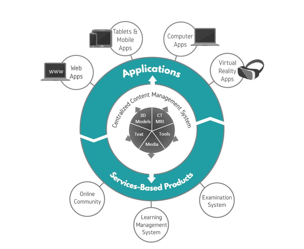
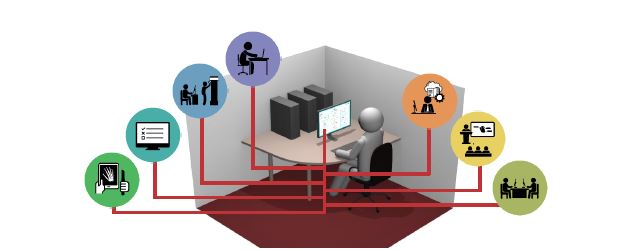
The graph in the next page shows how the components build up to form the solution. At the center, there are five star-shaped brackets that represent the five types of content generated by Forsina and the university. They enter the CMS, in which they’re sorted and edited, then assigned to Forsina products.
The first type of content is three-dimensional (3D), computer-generated (CG) models, created by a team of anatomists, doctors, and computer graphics specialists, to produce medical content digitally, in super quality and details. Forsina has created 3D CG models of all anatomy structures in the adult human male and female, and working to include the anatomy of the human child and embryos as well.
This extensive visual library includes over colored 30,000 structures in the human body, with superb surface details that are anatomically accurate and visually appealing, making this content an anatomical goldmine.
The superiority of 3D CG models is that they can be viewed from any angle, inside and out. Being digitalized, they can also be edited and dissected freely, in ways not possible or reversible with a real cadaver.
Forsina conducted its own CT and MRI whole-body scans on one human male and one female. The scanned persons are young, healthy and live individuals, providing a valuable medical reference of what live healthy human scans look like. It is rare to find a complete MRI scan for the same live individual, as you would find in Forsina’s unique content.
The scan data, converted into images, are grouped and implemented in many of Forsina application. CT images have seven pre-defined display modes, based on different tissue densities, to emphasize the visibility of certain tissues over others. All images have landmarks, which is a visual system for specifying the borders and names of apparent structures in each image.
Forsina has a team of doctors, anatomists, medical specialists, and content writers who gathered medical information from the most reliable references and combined them with years of accumulated medical experience to produce a massive medical encyclopedia that covers many essential subjects of health-sciences, like anatomy, histology, pathology, and biology.
In the anatomy section, every structure in the human body has a name and a detailed description. All of the 30,000 body structures are organized in a hierarchy with a special grouping system improvised by Forsina medical team.
A digital tool is an icon inside Forsina software with a specific pre-programmed function that is activated when the icon is clicked. Forsina has over 200 digital tools that grant users the ability to control Forsina 3D models, CT and MRI images, and the text encyclopedia, from within the applications.
Digital dissection tools are for dissecting and editing the 3D anatomical models, whether as a whole like making the human body lay down or stand up, or as parts like moving, isolating and enlarging certain structures.
Feature tools are for controlling important software features, like locating landmarks on bones surfaces and on CT images, or tracing blood circulation in a selected blood vessel, or video record students’ interaction with the digital body.
Multimedia content is a collection of millions of images, videos, and audio files, covering subjects of anatomy, histology, and medical imaging, produced solely by Forsina.
The images include numerous angle views for every anatomical structure among the 30,000 created by Forsina, and over 1,000 histology images.
The videos include 3D graphics explaining many subjects of anatomy, microbiology, and histology, as well as video recordings of anatomy lectures in universities. There are also informative videos, tutoring users on how to use Forsina products.
The audio library includes correct pronunciation of every structure, group, system, and landmark in anatomy, histology, and medical imaging, in multiple languages.
The original content management system. It has all the content created in productions for the last 10 years. New content is being added regularly and applied automatically to all related applications. The CMS enables distribution of new content to all applications, at the same, in a matter of clicks. It erases the pains of updating each application with new content manually.
The university uploads its data, like a CT scan of a cadaver, to a CMS replica that exists inside the university campus. This version is customized to the university’s needs and unique teaching model, and is synchronized with Forsina CMS. University's admins can perform the following functions on the CMS:
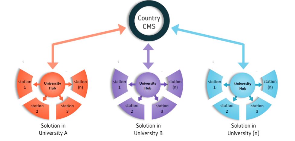
Every university using Forsina solution will have its own localized hub to control data flow and usage within its territory. The university has the option to connect its CMS to another university’s CMS, if they both agree to, to facilitate content transfer and sharing between the two institutions.
This version of CMS is meant to be used on a government level. It’s a centralized mega-control unit to control Forsina solutions in all member universities of a certain country. Usually, the ministry of higher education is in charge of this hub and uses it to oversee, regulate, and organize all content and software usage in the universities. Depending on regulations and law of each country, the university chooses to connect its hub to the country’s. Benefits include facilitating content exchange and data transfer, while the country ensures standards and guidelines are being adhered to among all members.
The country can connect its national hub with that of another country, based on a mutual agreement between them. Therefore, Forsina allows multi-levels of hub control; on an institution-scale, on a national-scale, and on an international-scale.
1-CMS: Content Management System
The CMS is a digital system for storing, organizing, and controlling all content and usage of Forsina software within the jurisdiction of the responsible institution. It has different tools and access privileges for the admin, professor, and student.
2-ES: Examination System
A system for making, managing and conducting examinations and quizzes inside the university. It provides exam-making tools for the professors, exam-analytics and management tools for the admin, and enable students to take examinations with an auto-grade feature.
1- Forsina Anatomy VR: Forsina Virtual Reality Application
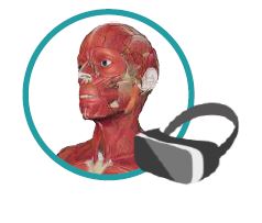
A virtual reality application teaching human anatomy and histology, with many dissection tools and features, designed for classrooms and group use. It offers a very high-quality 3D digital model of the male and female body and includes more than 30,000 body structures, 100 dissection tools, and an extensive library of medical text and images. It has exclusive advanced features like building Modules, tracing vessels, and shared VR group sessions.
2-Forsina Anatomy 3D: Forsina 3D Desktop Application
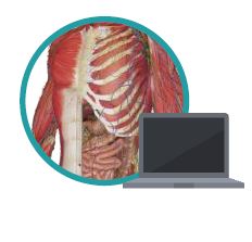
A computer application for teaching human anatomy and histology. It targets users with laptops or desktop computers. It has most of the features included in MVR, but without navigation in Virtual Reality.
1-Forsina Radiology VR
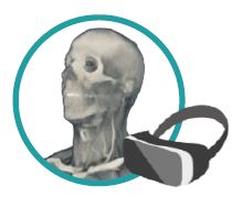
A Virtual Reality application teaching to read and understand Medical Imaging data, like CT and MRI scans, through 3D visualization and 2D images. It offers more than 500,000 CT/MRI images of healthy, live, young human adults, as well as scan for people with pathologies to serve as study cases. The images have more than 200,000 landmarks, accompanied by more than 100 software tools to edit the content, create new images, and record videos, all in virtual space.
2-Forsina Radiology 3D
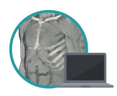
A computer application that teaches reading of CT and MRI data through 3D visualization and 2D images. It targets users of laptops and desktop computers. It offers the same 3D visualization in MiVR with the same features, except that it is viewed on computer monitors and controlled by regular mouse and keyboards.
1-Forsina Dicom converter VR
A Virtual Reality application that takes medical imaging data input from the user, in the .dicom file extension, and converts them into 3D models ready for navigation inside Virtual Reality. It provides tools to control the 3D model, explore inside it, and change tissue density to emphasize the visibility of certain tissues over others. Any CT/MRI scan data, of any patient, can be uploaded and explored. The application is a revolution in healthcare practice, serving as a very accurate diagnostic tool for medical doctors, and the perfect planning tools for surgeons to plan procedures before performing them.
The application is beneficial for education, allowing professors to upload Dicom files of people with different pathologies, and explore them with students as study cases.
2-Forsina Dicom converter 3D
A computer application that takes medical imaging data (Dicom files) from users and converts them into 3D models, viewed on monitors and navigated using regular mouse and keyboards. MiVR and MiDA are apps identical in functionality, but the first enables navigation in Virtual Reality as if one is examining the inside of a patient in real life, while the second allows this exploration on a computer screen.
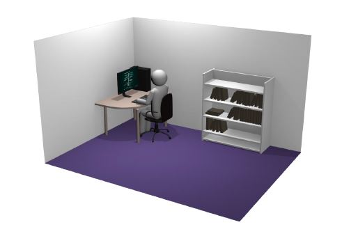
-The content control room is the core station, through which the university controls the whole solution.
The admin controls all the other stations using the CMS in the content control room.
-Activities:
Scan
The university conducts CT, MRI, or 3D scanning of new cadavers.
Upload
Upload scan data to CMS.
Convert
The CMS converts Dicom files to CT and MRI images, including images from three sectional planes, and seven different display modes to show seven pre-defined tissue densities.
Archive
Sorting and storing raw and converted data into corresponding directories.
Organize
Divide university users into groups with different privileges and access levels.
Assign
Update Forsina apps with university content to be edited and viewed by professors and students.
Distribute
Control which user groups get to view and edit which Forsina applications.
Station two is where professors prepare for anatomy lectures and dissection sessions.
Forsina enables professors to conduct the lecture in many ways. Whether by using PowerPoint presentations supported by visual aids, or dissecting a 3D digital body to explain anatomy in a practical approach, Forsina presents all of its educational applications of anatomy and medical imaging, in addition to the CMS, to professors, to make lecture preparing easier and more efficient. The applications offer:
1- An abundant source of 3D digital medical content.
2- A rich library of CT and MRI images
3- Text and images encyclopedia of anatomy and histology
4- Lecture-preparing tools.
Activities for preparing lectures:
Use Forsina Images
Use any number of medical images available in Forsina vast multimedia library, including millions of images of anatomy, histology, as well as CT and MRI scans.
Create New Images
Edit any of Forsina images and save the new files.
Copy Text
Use text from Forsina medical encyclopedia while prepping for the lecture.
Create Modules
Group 3D structures together that are related to the anatomy subject and save them in a new module.
Practice 3D Dissection
Practice doing digital dissections on MVR and MDA applications and explore the many advanced features provided to you.
Record Videos
Record yourself doing 3D dissection of the digital body or exploring the 3D holograms reconstructed from CT/MRI scans. Use the video recordings later in the lecture.
Create Exams and Quizzes
Using Forsina Examination System, you can create the examinations and assign them to students.
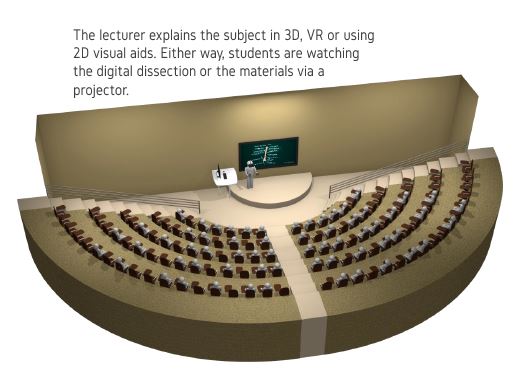
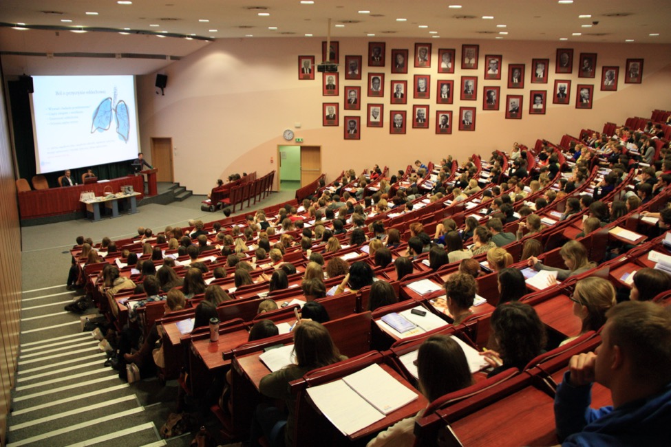
This station is where anatomy and medical imaging lectures take place. It is very beneficial to use 3D graphics and technological devices in this phase, because of the usually-large number of inexperienced students trying to understand complex medical subjects for the first time. Anatomy, for instance, is three-dimensional and it’s very important to comprehend the spatial relationships between parts and visualize their shape and color from all angles, that’s why using Forsina applications has never been more important.
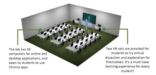
The digital room is a computer lab dedicated for students to study digital materials of anatomy, histology, and medical imaging, usually under supervision.
Activities in the digital lab:
Study
All materials shown in the lecture are available to students in the digital lab. All Forsina education applications are available on the desktop computers or the VR machines.
Experience Medical Virtual Experience
The VR machines allow students to experience a limitless and safe dissection. Whatever they saw the professors do in the lecture hall, they can do themselves in the digital lab.
Explore Anatomy and Medical Imaging in 3D
Students can explore 3D high-level medial content in MDA and MiDA, whether it’s 3D anatomy, or 3D holograms reconstructed from CT and MRI scans.
Peer Learning
Students can form groups and study together using Forsina applications. Peer learning accelerates and enhances education.
Ask Questions
VR machines allow the professor and student to join the same session and interact with the same 3D body, to ask questions and get answers promptly.
Save Notes
Notes can be added to any picture or on the 3D model, and saved locally or on CMS accounts.
Share
Any editing, questions, and notes can be shared with fellow students or with professors.
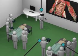
Station five is where cadavers are being dissected to show real human anatomy. Since any dissection action is irreversible and tissues and structures placements are ruined, Forsina software for virtual dissection is an important method to examine each structure from all angles and explain spatial relations between parts to students, before showing them on real cadavers.
This is where official university examinations are held. Forsina presents the examination system, Forsina ES, for universities to create, regulate and manage all exams and quizzes.
The system has three access gates for the university administrator, professor, and the student.
Admins are able to:
Professors are able to:
And Students can:
Receive assigned quizzes and submit them
The library is one of the most valuable information resources in the university. Forsina will provide its educational software for computers and tablets inside the library, and if possible, on VR machines too.
Activities in the library:
Study Anatomy in 3D
The superior software teaches all interested students about anatomy, using effective 3d graphics that make it easy to understand the subject, contrary to 2D images and text.
Study CT and MRI in 2D and 3D
Forsina allow students to check data in 2D and 3D in the library. Every image has many landmarks labeled and marked visually.
Take Pictures
The vast number of editable images available in the software is great for students to use in their presentations, research papers and study folders.
Conduct Research on Medical Subjects
Forsina applications are a valuable resource for students running research in subjects of anatomy and medical imaging, and even writing theses about the impact of employing technology in health sciences education.
Station Eight is the student’s home, where he/she studies by themselves.
Forsina is the student’s private teacher at home, teaching difficult medical topics, and providing a vast library of 3D medical content, images, text, and videos. Students are able to study from the very same materials used in the lecture and in the digital lab.
Forsina will provide the university with a number of single-user licenses. The licenses are to be distributed to students and used at home. Since students can rarely afford to buy Virtual Reality sets, only desktop computer apps are provided.
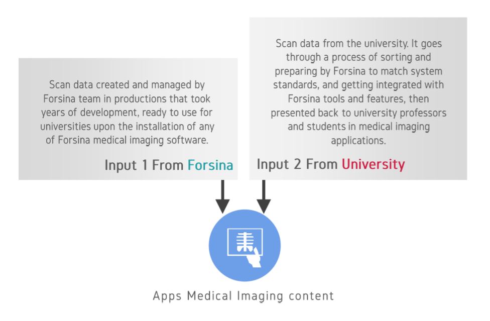
Forsina solution is flexible enough to use Forsina pre-made medical content and the university content inside the applications.
There are five types of content found throughout the solution. Most types accept input from Forsina itself and from the user. For example, when the content is medical imaging (MI) data (CT images, MRI images, Dicom files), the university can upload its own MI files, and the solution will process it and display it inside the applications. When using the educational applications, the end users (professors and students) can choose to view and edit Forsina MI content or the university’s MI content.
In other words, the university can combine the advanced functionality found in Forsina applications, with the university’s own content.
Available on Windows,macOS